For decades, observing real-time metabolic changes in deep brain structures has remained a major technical bottleneck in neuroscience research. Traditional imaging tools struggle to balance penetration depth and high-resolution cellular observation, leaving many brain metabolic mechanisms unsolved. Today, NADH brain metabolic imaging breaks this deadlock. As an endogenous fluorescent biomarker and core cellular energy molecule, NADH reveals previously invisible deep brain metabolic dynamics, opening a new chapter for precision neuroscience research and early neurological disease screening.
1. Why NADH Is the Ideal Marker for Brain Metabolic Research
NADH is widely recognized for its dual core functions in antioxidant defense and cellular energy metabolism. Beyond conventional health benefits, it possesses unique intrinsic fluorescent properties that make it an optimal label-free imaging marker for living brain cells. Its in-situ concentration and subcellular distribution directly reflect real-time mitochondrial energy status and cellular redox balance.
Slight fluctuations in brain NADH levels correspond to early physiological and pathological changes, including neuronal energy dysfunction, oxidative stress imbalance, and abnormal metabolic activity. These subtle alterations often appear long before clinical symptoms of neurological disorders such as Alzheimer’s disease, epilepsy, and neurodegeneration emerge. Therefore, precise visualization of NADH brain metabolic imaging enables researchers to capture the earliest warning signals of brain aging and neural lesions.
A 2025 breakthrough study published in Light: Science & Applications by Professor Osaki’s Japanese research team further elevates the application value of NADH brain metabolic imaging. The team developed a revolutionary label-free imaging technology, achieving high-resolution, deep-tissue observation of NADH in intact brain tissue for the first time, resolving long-standing limitations in neuroscience detection.
2. Technical Bottlenecks of Traditional Deep Brain Imaging
Brain tissue features dense cellular stacking and strong light scattering and absorption properties, analogous to a compact sponge structure. Traditional optical imaging techniques cannot penetrate deep brain layers, restricting in-depth neural metabolic research.
Conventional two-photon fluorescence imaging, though superior to one-photon imaging in penetration capacity, only reaches 100–200 micrometers, covering merely the superficial cerebral cortex. It fails to access core deep brain regions including the hippocampus and midbrain—the key areas closely associated with memory, cognition, and neural regulation.
Common clinical imaging methods such as ultrasound and MRI compensate for depth limitations but sacrifice cellular-level resolution. They can only display macroscopic tissue structural changes, unable to capture subtle metabolic fluctuations of single neurons and dynamic NADH distribution. For years, the lack of technology that balances “deep penetration” and “high cellular resolution” has hindered precise deep brain metabolic research—until the emergence of LF-MP-PAM technology centered on NADH brain metabolic imaging.

3. Revolutionary LF-MP-PAM: Core Principle of NADH Deep Brain Imaging
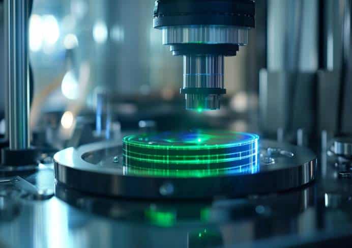
The newly developed label-free multiphoton photoacoustic microscopy (LF-MP-PAM) innovatively combines optical excitation and ultrasonic signal acquisition, perfectly solving the depth-resolution contradiction of traditional imaging. It relies entirely on endogenous NADH fluorescence for label-free detection, avoiding cell damage and exogenous marker interference.

3.1 Near-Infrared Femtosecond Laser: Ultra-Deep Optical Probe
The technology adopts a 1300 nm near-infrared femtosecond laser as the excitation source. Compared with visible light, this band features ultra-low tissue scattering and absorption, enabling efficient penetration into deep brain regions. Combined with a narrow 20–30 femtosecond pulsed three-photon excitation technique, it accurately triggers NADH fluorescence signals while preventing thermal damage to living neurons, ensuring safe and stable long-term dynamic monitoring.
3.2 High-Sensitivity Ultrasound Transducer: High-Definition Signal Capture
Laser-excited NADH produces subtle photoacoustic ultrasonic waves inside brain cells. The integrated high-sensitivity ultrasound transducer precisely captures these cellular acoustic signals. Since ultrasonic waves suffer far less attenuation in biological tissues than light waves, this method fundamentally breaks the depth limit of optical imaging. The system synchronously collects optical, photoacoustic, and third-harmonic signals for multi-dimensional verification, with high signal overlap ensuring highly accurate and reliable imaging results.
4. Breakthrough Imaging Performance: Ultra-Deep, High-Definition, Dynamic Monitoring
Driven by NADH brain metabolic imaging, LF-MP-PAM achieves industry-leading detection performance, comprehensively surpassing traditional imaging technologies:
Ultra-deep penetration capability: It reaches an imaging depth of 700 micrometers in mouse brain slices, over three times the penetration limit of traditional optical imaging. It clearly resolves subcellular NADH distribution in single neurons and accurately captures metabolic differences across brain regions—for instance, significantly higher NADH levels in the midbrain than in the cerebral cortex. In human brain organoid models, the maximum detection depth extends to 1100 micrometers, realizing unprecedented deep brain metabolic observation.
High-speed dynamic imaging: The system supports 256×256 pixel high-definition imaging at 0.76 frames per second, enabling real-time tracking of dynamic NADH metabolic changes. Experiments successfully capture rapid NADH elevation after 24-hour cellular hypoxia, perfectly matching known pathological metabolic rules and fully validating the practical reliability of NADH brain metabolic imaging.
5. Future Application Prospects: Reshaping Brain Health Research & Disease Prevention
This technological breakthrough is not limited to basic scientific research; it lays a solid foundation for future clinical precision brain health management and neurological disease intervention.
5.1 Early Screening of Neurological Diseases
Neurodegenerative diseases and epilepsy typically originate from early metabolic disorders, including neuronal energy deficiency and redox imbalance, which directly induce abnormal NADH metabolism. Traditional examinations can only detect structural lesions after symptom onset. In contrast,NADH brain metabolic imaging captures microscopic metabolic abnormalities at the pre-symptomatic stage, enabling ultra-early intervention and greatly improving clinical treatment outcomes.
5.2 In-Depth Exploration of Brain Development Mechanisms
NADH-mediated energy metabolism dominates fetal and infant brain development. This non-invasive label-free imaging technology allows real-time observation of deep brain metabolic dynamics, helping scientists reveal the core mechanisms of neural growth and differentiation, and providing new theoretical support for the prevention and treatment of pediatric developmental brain disorders.
5.3 Precision Personalized Brain Health Management
Lifestyle factors such as long-term staying up late, chronic stress, and fatigue directly disrupt brain NADH balance and trigger metabolic aging. In the future, NADH brain metabolic imaging can be applied to evaluate the impact of daily habits on brain health, achieving quantitative assessment of brain aging status and guiding personalized anti-aging and neuroprotection interventions.
6. Conclusion: NADH Decodes the Hidden Energy Code of the Brain
For a long time, human understanding of deep brain metabolism has been confined to superficial observations. The 2025 LF-MP-PAM imaging research redefines the value of NADH: it is not merely a cellular energy and antioxidant molecule, but a core biological code for deciphering deep brain metabolic mysteries.
Through high-precision, ultra-deep NADH brain metabolic imaging, humans can finally directly observe real-time energy operation and redox changes in deep brain neurons. This breakthrough opens a new era for neuroscience research, early neurological disease warning, and precise brain anti-aging, bringing unlimited possibilities for future human brain health protection.
References
Osaki T, Lee W D, Zhang X, et al. Multi-photon, label-free photoacoustic and optical imaging of NADH in brain cells[J]. Light: Science & Applications, 2025, 14(1): 264.